


Thirteen years ago, microbiologist Christa Schleper and her coworkers hauled a tube of mud from the floor of the North Sea. In it they discovered a group of microbes that could tell us about the origins of our branch in the tree of life. Now they’ve finally managed to grow enough in the lab that they can look at them under a microscope. Here’s how—and why—they did that.
“Asgard archaea are considered to be the closest known relatives of eukaryotes.1 Their genomes2 contain hundreds of eukaryotic signature proteins (ESPs),3 which inspired hypotheses on the evolution of the eukaryotic cell.4 A role of ESPs in the formation of an elaborate cytoskeleton and complex cellular structures has been postulated,5 but never visualized…6
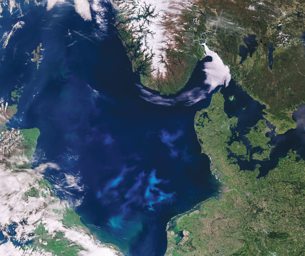
Considering that lokiarchaeal organisms and other Asgard archaea can be found in a variety of anoxic and often marine environments, we screened DNA from shallow-water sediment from different locations for the presence of 16S rRNA genes of Asgard archaea to select suitable and easily reachable sampling sites for establishing enrichments.7 Sediments from a small estuarine canal that regularly receives water from the Mediterranean near the coast of Piran, Slovenia, were identified to have the highest relative abundance at the 13–16 cm depth layer8, exhibiting up to 4%9 of Asgard archaea 16S rRNA genes in amplicon sequencing…
With periodic monitoring using quantitative PCR (qPCR) with Lokiarchaea-specific primers, growth could be observed after 140 days at 20 °C10 in serum flasks containing sterile-filtered water from the original source supplemented with complex organics (casein hydrolysate, milk powder and amino acids). However, after two transfers under these conditions, growth could no longer be detected and a second round of screening with different medium compositions was performed.11 Using a modification of the medium MK-D1 reported for the cultivation of ‘Ca. P. syntrophicum,’ cell growth recovered, and abundances reached repeatedly 2–8%. However, higher enrichments were not achieved under these conditions. Only through developing a minimal medium, mostly by reducing the input of organic carbon sources to a single compound and by increasing antibiotic concentrations, lokiarchaeal relative abundances reached between 25% and 80% after several transfers.…12
Amplicon sequencing analyses of 16S rRNA genes13 revealed that the culture with the highest enrichment (Loki-B35) consisted of three dominant and two minor species14: a single Lokiarchaeon sequence (79%), a sulfate-reducing bacterium of the Desulfovibrio genus (10%), a hydrogenotrophic methanogen of the Methanogenium lineage (6%), as well as a Halodesulfovibrio and a member of the Methanofastidiosales genus (both at around 2%).…
We next plunge-froze cells of a live culture onto electron microscopy (EM) grids to image them in a near-native state using cryo-electron tomography (cryo-ET).…15 This approach revealed three general cell types that had distinct morphologies and cell envelope architectures. One class consisted of round-shaped cell bodies associated with elaborate and heterogeneous protrusions.16 The other two cell types were rods and spherically shaped cells,17 respectively, without protrusions. Their cell envelopes had canonical Gram-negative and archaeal cell envelope features, therefore probably representing co-cultured bacteria and archaea. By contrast, the cell envelope of ‘Ca. L. ossiferum’ candidates featured complex unordered densities18 protruding from a single membrane.…
Having established the identity and general appearance of ‘Ca. L. ossiferum’ cells, we next aimed to analyse their overall organization by scanning EM. We identified small coccoid cells with surface-bound vesicles and extensive protrusions. In contrast to ‘Ca. P. syntrophicum,’ these long protrusions appeared more irregular, frequently branching or expanding into bulbous structures…19
We discovered an elaborate actin-based cytoskeleton in Asgard archaea,20 which has long been hypothesized, but has not been visualized…
The elaborate cell architecture with extensive membranous protrusions has multiple implications for Asgard physiology and ecology. As these characteristic features make the cells highly fragile,21 it could also explain why the highest abundance of Asgard archaea is found in sediments rather than in plankton.…
The large surface area of the convoluted network of protrusions, in combination with the unusual cell envelope lacking a highly ordered S-layer (as typically found in other archaea) but rather displaying numerous surface proteins, may have enabled the intricate cell–cell contacts required for eukaryogenesis that—considering the lifestyle of the two cultured Asgard strains—probably involved interspecies dependencies in syntrophic relationships. These findings strongly support a gradual path of mitochondrial acquisition through protrusion-mediated cell–cell interactions.22”
*
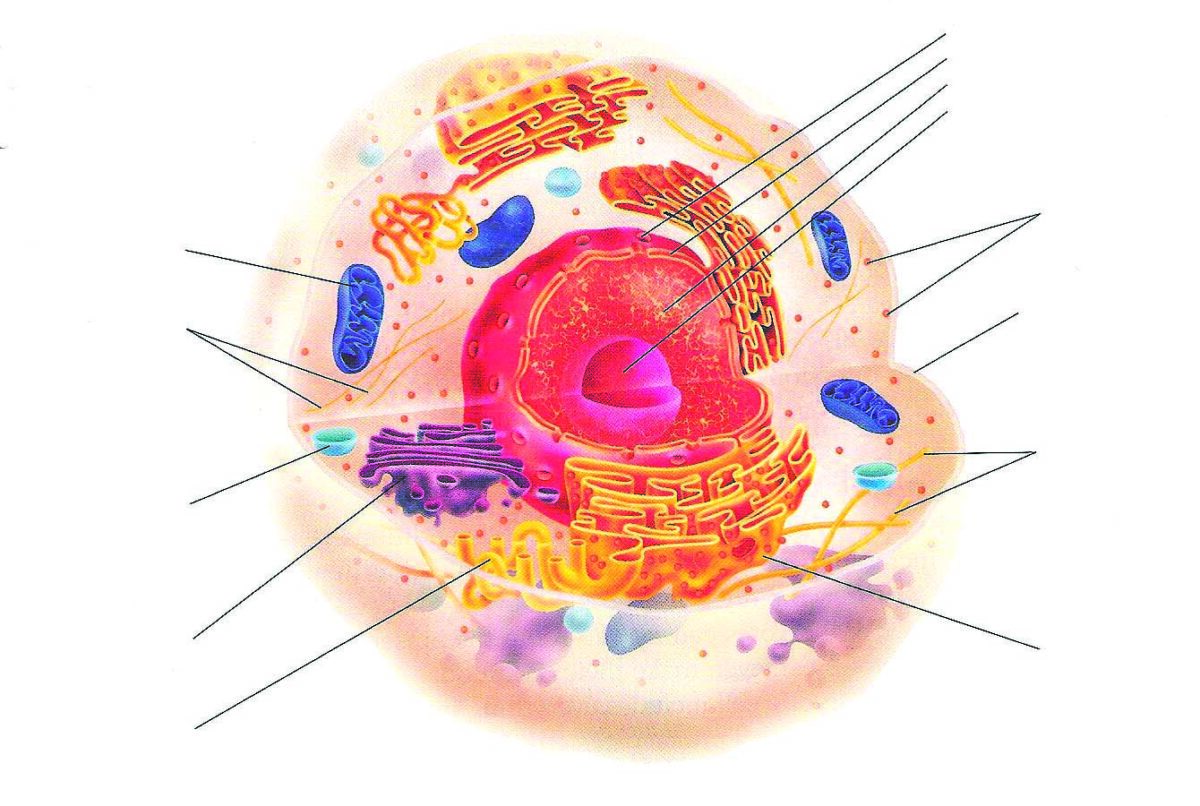
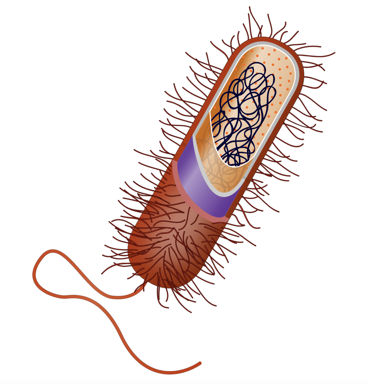
1 First, the basics: You are a eukaryote. So is everything whose cells have a nucleus and mitochondria. Bacteria are not; they’re prokaryotes, and somewhere way back, it was a prokaryote that evolved and changed and became the first eukaryote. Mitochondria are the remnants of a long-ago bacterium that was somehow subsumed by early eukaryotes. Archaea are neither bacteria nor eukaryotes. They are microbes, but aspects of their cells seem to be like ours. That’s especially true of the Asgard archaea, the ones Schleper and her colleagues detected for the first time in that North Sea mud.
2 Important note: Asgards’ genomes, which you can fish out of the mud without culturing a living organism, are almost the only thing researchers have been able to study so far. These bugs are wildly finicky about their living situation: they are the hothouse orchids of the microbial world.
3 These are genes that we thought only eukaryotes had, things that make us special. Until the Asgards came on the radar.
4 That is to say: Are they a peek into what our earliest eukaryotic ancestors might have looked like?
5 Some of these genes code for a protein that looks like actin, the building block of a kind of scaffolding inside your cells. Are Asgards structuring their cells like we do?
6 An understatement! Before this paper, the only time anyone had actually seen an Asgard archaeon was after a Japanese group spent twelve years culturing enough of them to photograph. They got only a few snapshots, and they didn’t get to do experiments with the cells before the cells died
7 They looked in a lot of places for some nice high-quality mud.
8 This Slovenian ditch had the highest density of Asgard archaea of anywhere they looked. If you’re going to grow them in the lab and have enough to do experiments with, you have to start with an Asgard-heavy sample.
9 Four percent doesn’t sound like much, but that’s a lot of Asgard archaea in one place!
10 They’re growing! Exciting!
11 They stopped growing. Sigh.
12 It took months and months of trying lots of different things for the scientists to get these Asgards—a type known as Lokiarchaea—to grow well. They were trying various tricks that the Japanese group had used, which included Japanese baby formula.
13 Before they tried to look at the cells, they did DNA testing to see what exactly had survived.
14 What they found: These Asgards grow best when they have some bacterial friends living with them. The researchers think these bacteria may be producing molecules that Asgards can eat, providing a kind of neighborly buffet.
15 To take a look at the cells, they froze the Asgards and their bacterial friends, slivered them like fine pastrami, then bounced rays of electrons off them. This produces crisp images of the cells’ silhouettes.
16 These are the Asgards.
17 These are the bacteria.
18 My new band name. Also, a way of saying that these archaea are funny-looking.
19 Once they finally got to see the archaea, they were wildly odd. They had snaky arms, little tentacles, strange blobby bits.
20 What’s more, the scientists were able to confirm that the Asgard cells had something forming threads within them, like the actin in your cells, confirming their strange synchrony with eukaryotes.
21 These wiggly bits are very delicate, helping to explain why, until Schleper and her colleagues pulled up their North Sea mud, nobody had noticed Asgards living all around us in the soil—touch them and they fall apart.
22 And here, folks, is the mic drop: If Asgards have long limbs that they can drape over their generous bacterial neighbors, could it be that we’re seeing something like what happened when early eukaryotes picked up the bacterium that became the mitochondrion? Did early eukaryotes extend a protrusion and start a chain of events that led to you, here, now, reading this page?